-
The neuronal basis of self-motion perception
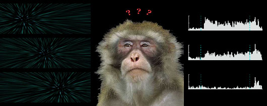
When we move through the natural scene, the image on our retina is also in motion. Gibson termed this self-induced visual motion "optic flow". Optic flow provides visual input to guide our self-movement. If we keep our eyes still when we move forward, the retinal image appears to expand. The center of this expansion pattern indicates our self-movement direction, which we can also call heading direction.

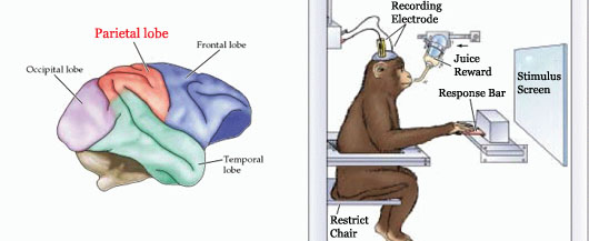
High-order areas in the visual motion system of dorsal extrastriate cortex have been implicated in the analysis of optic flow stimuli that helps to support perception of observer heading. For the past few years, we have been using the ventral intraparietal area (VIP, located at the bottom of intraparietal lobe) as a model for studying how optic flow information is processed and the functional role of VIP in heading direction discrimination task.

Monkeys are trained to perform demanding perceptual tasks, and we record from single or multiple neurons in cortex during performance of these tasks. This allows us to directly investigate the neuronal mechanism underlying particular perceptual behavior. In addition, Electrical microstimulation is used to test the direct linkage between physiology and behavior. Besides the standard biostatistical methods, we also use computational modeling to interpret results and guide future studies.

